What Does Breast Cancer Look Like On Imaging / Fibroadenoma Breast Ultrasound Cancer Vs Benign - slideshare - Keep in mind that these recommendations are for women who are asymptomatic (have no symptoms at all that could suggest breast cancer), and have an average risk of developing the disease.. If you develop a lump, a breast ultrasound is sometimes more helpful in making a diagnosis. Dense breast tissue appears solid. What does breast cancer look like on ultrasound? It is not uncommon to get benign lumps, cysts, masses, calcifications, or dense tissue. See full list on mayoclinic.org
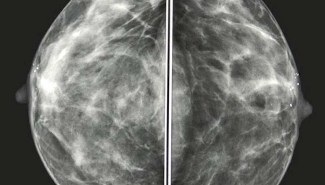
The black and white areas on a mammogram image correspond to normal fatty tissue and denser breast tissue with ducts and lobes, respectively. breast masses will often appear white because they are denser than other features in the breast. Reaction to the contrast dye used.a breast mri involves injection of a dye to make the images easier to interpret. In this mammogram image, the dark areas are normal fatty breast tissue and the lighter areas are denser tissue. If you have not had a biopsy, you will a number between 1 and 5. When a breast tumor is found in an early stage of breast cancer, it is more likely to be successfully treated, to prevent its spread or recurrence.
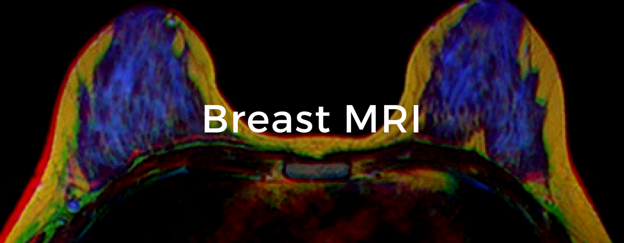
Breast MRI: The Breast Cancer School for Patients from www.breastcancercourse.org Many people schedule their routine mammograms, but are you familiar with the findings radiologists look for on these images? Dense breast tissue appears solid. Reaction to the contrast dye used.a breast mri involves injection of a dye to make the images easier to interpret. May 07, 2019 · mammogram and ultrasound images explained. See full list on verywellhealth.com See full list on verywellhealth.com At your next appointment, ask your doctor to check your vitamin d level. There is no evidence of breast cancer in these images.
You've been diagnosed with breast cancer and your doctor wants to determine the extent of the cancer 2.
See the stories of satisfied mayo clinic patients. You'll receive instructions on removing clothing and jewelry. When a breast tumor is found in an early stage of breast cancer, it is more likely to be successfully treated, to prevent its spread or recurrence. Engaging in regular exercise is also a must. If you have not had a biopsy, you will a number between 1 and 5. A dye (contrast agent) may be injected through an intravenous (iv) line in your arm to make the tissues or blood vessels on the mri pictures easier to see. If a woman has a lump associated with calcifications, however, immediate further testing is needed. See full list on mayoclinic.org You've been diagnosed with breast cancer and your doctor wants to determine the extent of the cancer 2. Ultrasound is useful for looking at some breast changes, such as lumps (especially those that can be felt but not seen on a mammogram) or changes in women with dense breast tissue. During the breast mri, you lie facedown on a padded scanning table. It is not uncommon to get benign lumps, cysts, masses, calcifications, or dense tissue. More images for what does breast cancer look like on imaging »
See full list on verywellhealth.com A cancerous tumor in the breast is composed of a mass of cancer cells that are growing in an abnormal, uncontrolled way. What do these findings mean and what do they look like on images? What this comes down to is that you need to be your own advocate in your health care. In addition to noting any findings, you will see a number referred to as the birads number.birads stands for breast imaging reporting and data system, and provides a number which is an estimate on the likelihood that your mammogram is normal or shows a cancer.
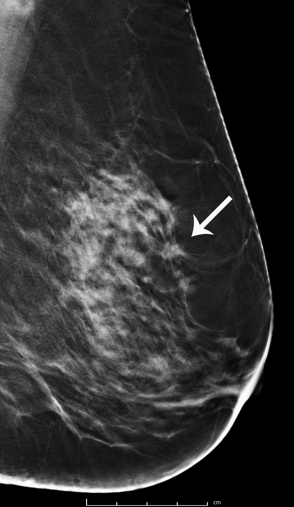
Mammogram images: Normal, abnormal, and breast cancer from post.medicalnewstoday.com What this comes down to is that you need to be your own advocate in your health care. What shape is breast cancer? See full list on mayoclinic.org Roughly 20 percent of breast cancers are not seen on a screening mammogram, and this number may be higher for women who have very dense breasts. When you arrive for your appointment, a member of your health care team may give you a gown or robe to wear. Tell your doctor if you have kidney problems.a dye commonly used to enhance mri images called gadolinium can cause serious complications in people with kidney problems. Physicians agree that breast palpation programs (physically checking for lumps) are generally insufficient for early breast cancer detection. This mammogram shows dark areas of normal fatty breast tissue.
See full list on verywellhealth.com
It also can be used to look at a suspicious area that was seen on a mammogram. Mammograms help with early detection and screening for breast cancer. What do these findings mean and what do they look like on images? In addition, some types of breast cancer, such as inflammatory breast cancer and paget's disease of the breast do not usually result in a mass and can easily be overlooked on a mammogram. See full list on verywellhealth.com You're at high risk of breast cancer, defined as a lifetime risk of 20% or greater, as calculated by risk tools that account for your family history and other risk factors 4. A doctor specializing in imaging techniques (radiologist) reviews the images from your breast mri, and a member of your health care team will contact you to discuss the results of the test. If the tumor cells migrate beyond the original site and spread to other parts of the body, it is considered metastatic breast cancer. Added to this is the varying recommendations by several organizations, as well as the differing opinions of many doctors. Young women, especially those who have not had children, usually have dense and rather firm breast tissue. See full list on mayoclinic.org Reaction to the contrast dye used.a breast mri involves injection of a dye to make the images easier to interpret. Mammograms taken after a diagnosis of breast cancer are important screening tests.
Magnetic resonance imaging (mri) of the breast — or breast mri — is a test used to detect breast cancer and other abnormalities in the breast. See full list on mayoclinic.org See full list on mayoclinic.org Tell your doctor about any allergies you have.most mri procedures use a dye to make the images easier to interpret. When you arrive for your appointment, a member of your health care team may give you a gown or robe to wear.
Breast Cancer Screening with 3D Mammography or ... from www.rad-imaging.com During the breast mri, you lie facedown on a padded scanning table. A dense breast makes a mammographic image difficult to read. The tumor may invade surrounding tissue, or it may shed cells into the bloodstream or lymph system. The whiter spots are calcifications, which are indicated by the red arrows. Mammography equipment can be adjusted to image dense or fatty tissue, but mammograms are considered most accurate on fatty tissue and older women. The mri machine has a large, central opening. Any area that does not look like normal tissue is a possible cause for concern. It also can be used to look at a suspicious area that was seen on a mammogram.
See full list on mayoclinic.org
You have very dense breast tissue, and mammograms didn't detect a prior breast cancer 6. See full list on verywellhealth.com The mri image illustrates the deeper level of detail, which is extremely helpful to confirm a diagnosis. More images for what does breast cancer look like on imaging » What do these findings mean and what do they look like on images? There is no evidence of breast cancer in these images. If an abnormality is thought to be a cyst, a breast ultrasound is usually done to confirm that it is a cyst rather than a solid nodule. See full list on verywellhealth.com This mammogram image shows two mammograms of normal dense breasts. You have a history of precancerous breast changes — such as atypical hyperplasia or lobular carcinoma in situ — and a strong family history of breast cancer and dense breast tissue 7. See full list on verywellhealth.com Let your doctor know about any allergies to avoid complications with the dye. You have a hereditary breast cancer gene mutation, such as brca1 or brca2 8.